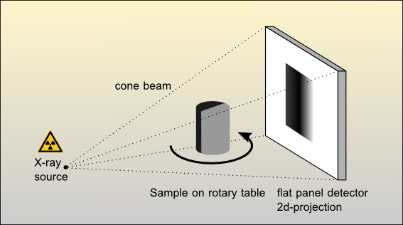
In material sciences computer tomography (CT) is a non-destructive method for the 3D (4D) characterization of materials. The general setup for CT scans consists of a “light” source, a sample on a turntable and a detector. In a CT scan, the sample is stepwise rotated by 360° (180°) and at each step the picture (2D) on the detector is recorded. A 3D model of the sample is computed based on this data. If the acquisition time of a scan is fast enough, the sample can also be studied in 4D. Usually X-rays, synchrotron radiation or neutrons are used as the light source for CT scans.
Currently the CT lab is equipped with a high resolution X-ray computed tomography (HRXCT)-scanner. HRXCT, or µ-X-ray Computed Tomography, visualizes and quantifies features of X-ray transparent materials (solid and liquid) and produces images that correspond closely to serial sections through the sample. These images reflect the variation of the X-ray attenuation, which is mainly a function of the density, within the sample. Sectional contiguous images are combined to generate a 3D model of the density distribution. This model is used further for visualization and quantification of the geometrical characteristics of the sample (e.g. pore volume, phase abundances, spatial distribution of phases, grain size distributions etc.). For more information about HRXCT scanning techniques seehttp://www.ctlab.geo.utexas.edu/overview/index.php.
The micro computer tomography laboratory at the Institute of Geoscience at the University of Mainz is financed by the excellence cluster “geocycles – time and space in the earth sciences” of the federal state Rheinland-Pfalz, the DFG (DFG INST 247/519-1) and by the Johannes Gutenberg University of Mainz. The laboratory provides HRXCT and post-processing of data for in-house users, external collaborators and for external users.
Terms for the laboratory usage are subject to negotiation and requests should be sent to Frieder Enzmann. |